
Electroencephalography (EEG) is the measurement of electrical patterns at the surface of the scalp which reflect cortical activity, and are commonly referred to as “brainwaves”.
 An important assessment tool is an EEG brain map, also called QEEg or Quantitative EEG. It helps give us a picture of what’s going on in the brain. There are many different types of brain maps – including MRI’s, PET scans, and SPECT scans. But the EEG map provides the best information for neurofeedback training. It shows brain timing issues which impact mood, behaviour, and attention.
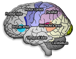
An EEG brain map helps identify where the brain has specific problems – and helps target the kind of interventions. For neurofeedback, it provides a guide to where to train. Each area of the brain plays an important functional role. If one or more areas of the brain is running too slowly or too fast, it causes problems – such as attention, emotional control, mood or behaviour difficulties. One area of the brain is constantly communicating with other areas. If they are not communicating well, it will interfere with learning and create other problems.
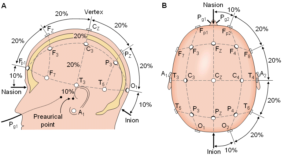
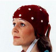
These measurements can be done at various sites on the scalp (according to the 10-20 site map). This can be done with individual electrodes or electrodes imbedded into an electro-cap.
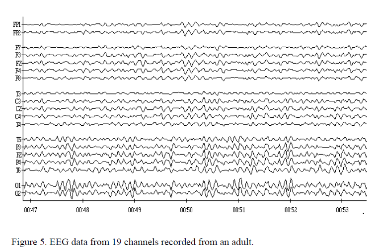
This image is of 19 channel EEG recording done digitally on a computer. These recordings will firstly be visually interpreted to get an understanding of the general EEG and brain function patterns. This is not used as a diagnostic tool in the hands of the Neurotherapist but rather in giving understanding and guidance for training towards better regulated brain patterns. Neurologists will be best able to interpret for dysregulations like Epilepsy or any other pathology.
These digitised recordings can then be processed and analysed using various algorithms such as the Fast Fourier transform or comparing values with a normative database – this process produces a Quantitative EEG or also called Brain Map.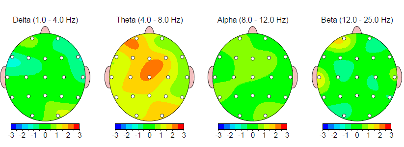
The EEG and the now processed QEEG information can be interpreted and experts in the field use it as a clinical tool to evaluate brain function and to look for changes in brain function during and after interventions like Neurofeedback, Mindfulness Stress reduction or other mainstream therapeutic processes, even medication effect.
This Mapping process gives us the ability to observe dynamic changes in the brain when the brain is engaged in for instance cognitive tasks - showing brain areas that are not effectively engaging or processing information as the task demands.
These Brain Maps form part of the clinical evaluation and data acquisition of the Neurotherapist in order to effectively draw clinical conclusions and construct an individualised treatment plan for the client.
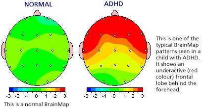
The example above is of a client with ADHD symptoms. The frontal lobes shows excessive immature and slow Theta wave activity (red). This can be corrected with medication or Neurofeedback training or both.
Results of assessments / tests as well as clinical observations of behaviour patterns together with the brain map findings lead to more effective brain function conclusions. These findings guides the Neurotherapist in using research based protocols to train towards better regulated brain and nervous system regulation. The outcome is then more effective daily functioning.